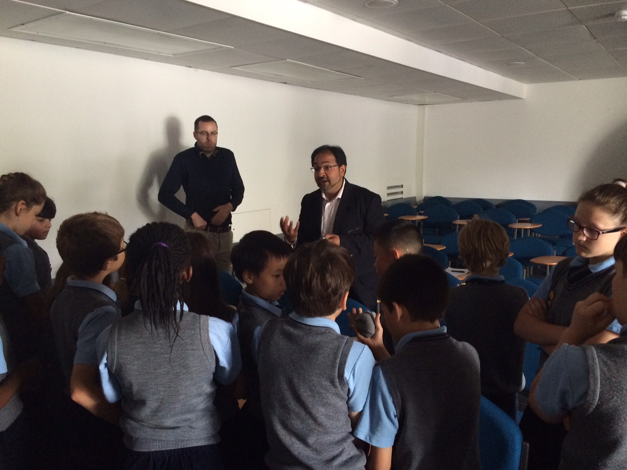
Dr Christina Malamateniou is a Lecturer of Perinatal Imaging in the Department of Perinatal Imaging and Health, part of the Division of Imaging Sciences & Biomedical Engineering. She organised a morning of talks and activities, showcasing King’s top quality clinical research to a local primary school.
There has never been a more exciting time for children to engage with the latest clinical science developments than now: new, more powerful and sophisticated MRI scanners are being built, a multitude of advanced imaging techniques are being developed-to study normal development and disease as early in the course of life as possible, starting (literally) from the womb(!); 3D-printing and robotics have brought visualisation and understanding of complex pathologies to a totally new level and genetics promises to disentangle the mysteries of human life!
My colleagues at King’s College London and I are passionate about both our clinical research and science and it was exactly this passion that we wanted to instil to 25 ten-year-olds from Heath House Prep School when they visited us at King’s College London Waterloo Campus at the end of summer 2015.
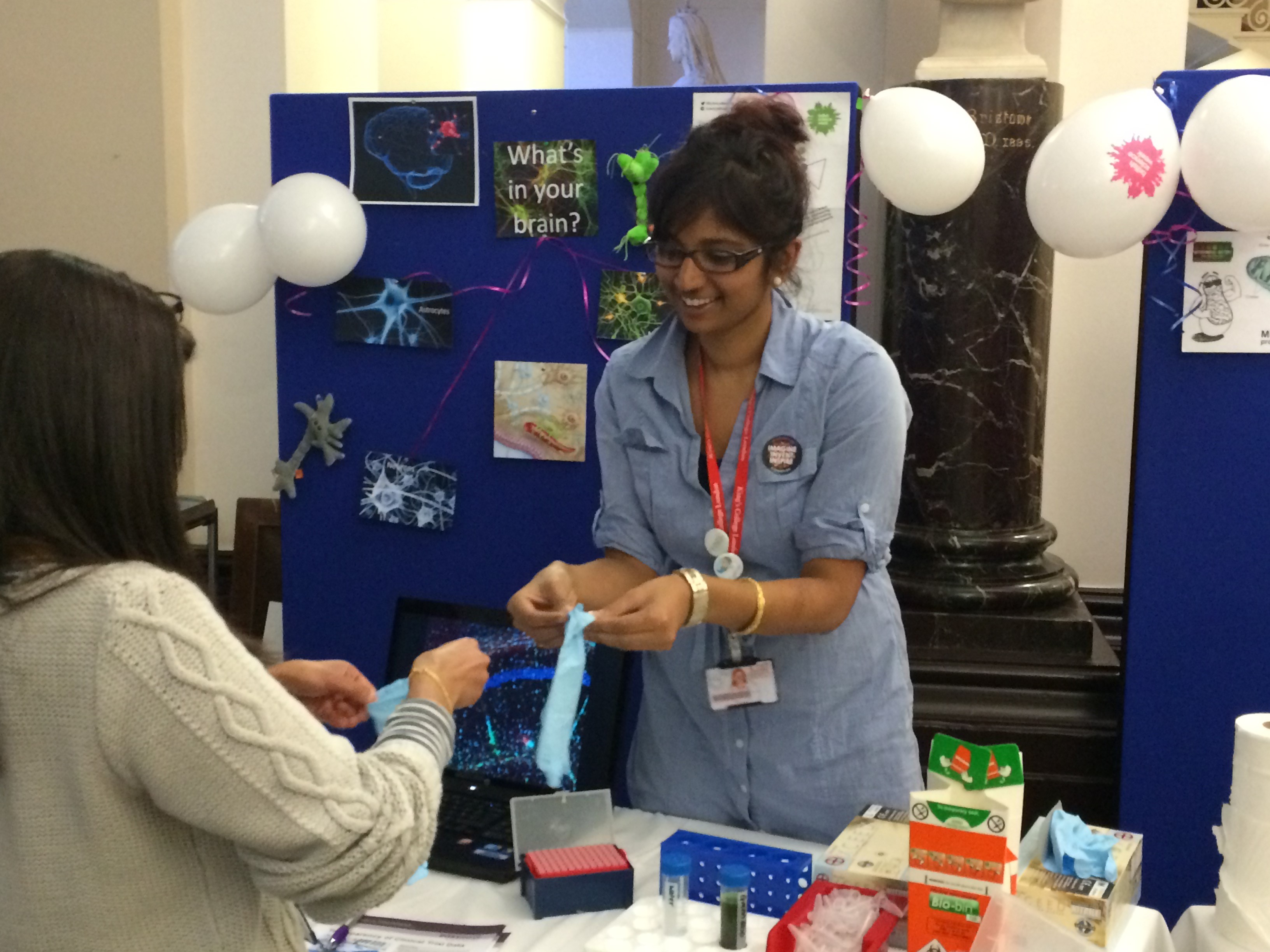
It was a  pleasant morning at the Franklin Wilkins Library and I started our day describing how MRI scans are done in our department (Perinatal Imaging and Health) and how the clever imaging we use can make a difference in the quality of life of the very premature babies; children giggled when they saw for the first time how a fetus moves inside his mummy’s tummy and they had the chance to play with an inflatable full size MRI scanner too (a.k.a. the Giant Doughnut, “wow, now I can see why it makes such a loud noise, it is huge!”, kids remarked during the break).
 The children then got a chance to learn cool facts about the most amazing, tireless muscle of our body: the human heart and got excited with the robotic arms that Dr Kawal Rhode and his PhD student Shuangyi Wang have constructed to study it. During the break kids saw, and even hold, 3D printed copies of human hearts and they got to guess which one belonged to a premature baby!
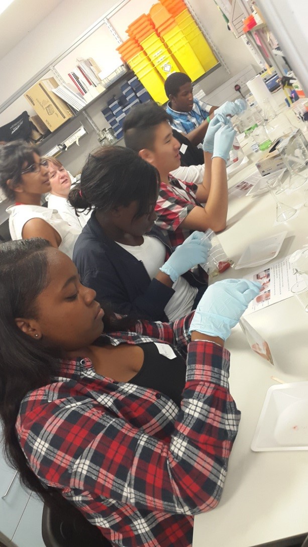
 Dr Claire Thornton took the baton to introduce us to the magic of genetics and what better way than helping the children learn how to extract DNA from the most fragrant fruit of the summer: strawberries. The students were absolutely thrilled with all the lab equipment they got to use and they amazed us with their questions and eagerness to learn more. We could clearly see some little scientists in the making there!
After an inspiring morning, the children were introduced to our library facilities and academic study during a fun-packed event, carefully planned and organised by our amazing library staff. Mrs Beata Gędłek and her wonderful team navigated the children in the library quiet zones, discussion rooms, study pods (with kids commenting that they looked like coming out of a star wars movie!) and they organized a fun book treasure hunt, with books relating to the morning talks. Kids were divided into 5 groups (Medicine, Biology, Genetics, Robotics and Physiology) and they were like little science detectives as they looked for the specified books using maps and clues carefully prepared by the library staff. They then presented their results and answered questions in the high-tech touch-screen newly installed computer room in the library. There were all smiles when they were awarded their library super-user certificates and KCL gifts at the end of the day.
What an eye opening experience for the children and great fun for us too; kids were asking the most amazing questions, participating and engaging with science, their eyes lighting up with enthusiasm. They were clearly overwhelmed by the amazing facts they learned during our fun interactive workshop. Their feedback says it all: “That was the most amazing visit of our school this year”, “I learned about babies born early and strawberry DNA”, “I didn’t want the day to end!”, “I think I want to become a scientist after all”.
 The success of this event and the smiles on children’s excited faces was the best reward for us all. It was also a great way to advertise the multidisciplinary top quality research that takes place everyday at King’s College London across many different departments. I am indebted to all the people who enthusiastically came along and shared their passion in doing research and teaching science to these young inquisitive brains. I guess for us all there is only one question: “When is the next time we can do it again?!”